
Файл:Mycobacterium tuberculosis.jpg
Төп файл (1997 × 1927 нокта, файл зурлыгы: 543 Кб, MIME төре: image/jpeg)
Тасвирлама
| Тасвир |
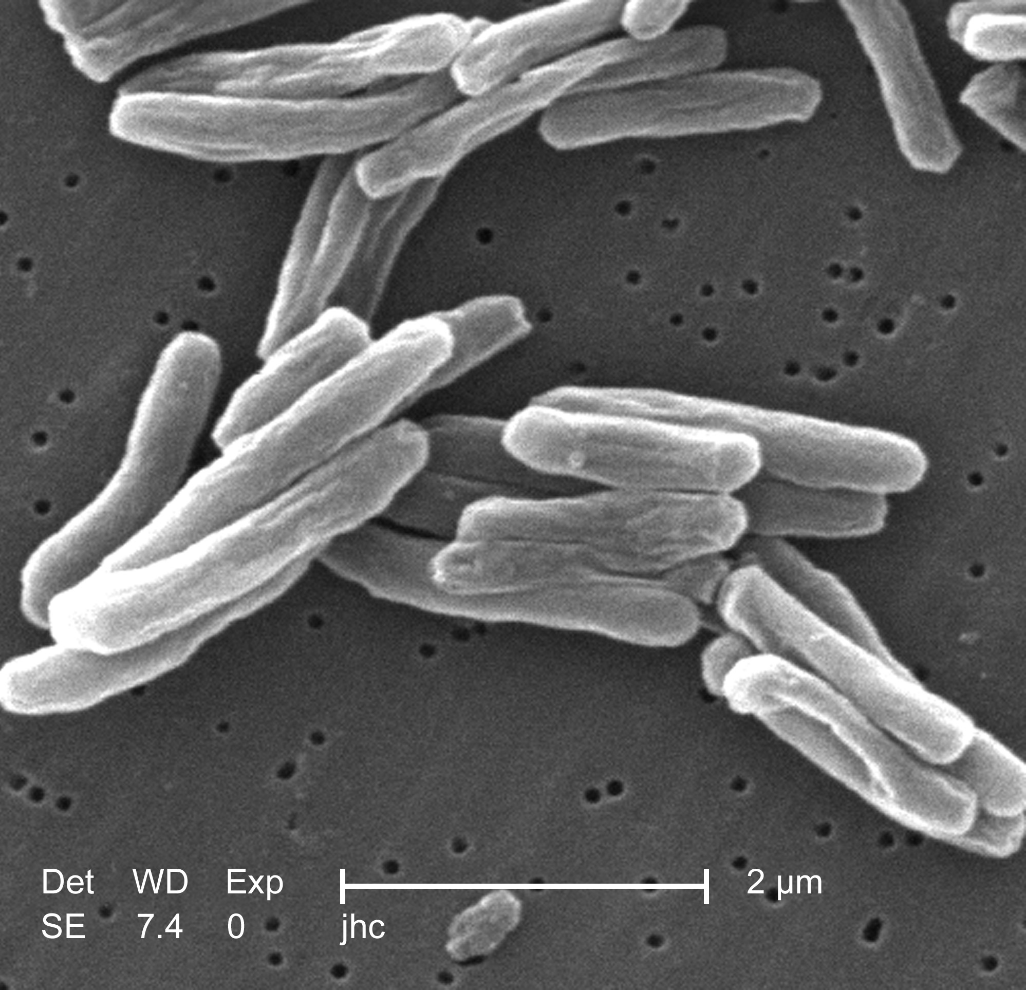
English: Under a high magnification of 15549x, this scanning electron micrograph (SEM) depicted some of the ultrastructural details seen in the cell wall configuration of a number of Gram-positive Mycobacterium tuberculosis bacteria. As an obligate aerobic organism M. tuberculosis can only survive in an environment containing oxygen. This bacterium ranges in length between 2 - 4 microns, and a width between 0.2 - 0.5 microns. See PHIL 9997 for a colorized version of this image.
TB bacteria become active, and begin to multiply, if the immune system can't stop them from growing. The bacteria attack the body and destroy tissue. If in the lungs, the bacteria can actually create a hole in the lung tissue. Some people develop active TB disease soon after becoming infected, before their immune system can fight off the bacteria. Other people may get sick later, when their immune system becomes weak for another reason. Babies and young children often have weak immune systems. People infected with HIV, the virus that causes AIDS, have very weak immune systems. Other people can have weak immune systems, too, especially people with any of these conditions: substance abuse; diabetes mellitus; silicosis; cancer of the head or neck; leukemia or Hodgkin's disease; severe kidney disease; low body weight; certain medical treatments (such as corticosteroid treatment or organ transplants); specialized treatment for rheumatoid arthritis, or Crohn's diseaseFrançais : Mycobacterium tuberculosis grossi 15 549 fois.
Español: Mycobacterium tuberculosis ampliado a 15549x.
中文:掃描電子顯微鏡下的結核桿菌.
Suomi: Mycobacterium tuberculosis 15549-kertaisena suurennoksena.
Čeština: Bakterie Mycobacterium tuberculosis, původce TBC.
Magyar: Mycobacterium tuberculosis.
한국어: 결핵균의 전자현미경 사진.
Simple English: TB Bacteria.
Kurdî: Girtineke elektronmîkroskobîk a bakteriyên tûberkûlozê pêk tînin.
Afrikaans: 'n Skanderende mikrograaf van Mycobacterium tuberculosis.
粵語: 掃描電子顯微鏡下嘅結核桿菌. |
||
| Дата | |||
| Чыганак |
|
||
| Автор |
|
||
| Рөхсәт (Бу файлны кабат куллану) |
PD-USGov-HHS-CDC English: None - This image is in the public domain and thus free of any copyright restrictions. As a matter of courtesy we request that the content provider be credited and notified in any public or private usage of this image. |
||
| Башка юрамалар |
|

Лицензияләү
Это изображение было создано или получено агентством Центры по контролю и профилактике заболеваний, которое является подразделением Министерства здравоохранения и социальных служб США. Это было сделано при выполнении служебных обязанностей государственным служащим. Поскольку изображение создано Федеральным правительством США, оно находится в общественном достоянии (public domain).
|
Исходный журнал загрузок
- 2006-10-06 23:04 TimVickers 220×212×8 (10514 bytes) Janice Carr, CDC, http://www.cbc.ca/health/story/2006/03/17/tb-who060317.html?ref=rss
Файл тарихы
Файлның нинди булганлыгын күрү өчен «дата/вакыт» дигәненә басыгыз.
| Дата/вакыт | Кече рәсем | Үлчәмнәре | Кулланучы | Искәрмә | |
|---|---|---|---|---|---|
| агымдагы | 21 июн 2019, 15:31 | | 1997 × 1927 (543 Кб) | Tholme | Higher resolution |
| 9 июл 2008, 00:53 |  | 220 × 212 (10 Кб) | Reborned | {{Information |Description={{en|Janice Carr, CDC, http://www.cbc.ca/health/story/2006/03/17/tb-who060317.html?ref=rss}} |Source=Transferred from [http://en.wikipedia.org en.wikipedia] |Date=2006-10-06 (original upload date) |Author=Original uploader was [ |
Файлны куллану
Әлеге файл киләсе битне куллана:
Файлның гомуми кулланышы
Әлеге файл аста бирелгән викиларда куллана:
- af.wikipedia.org проектында куллану
- ang.wikipedia.org проектында куллану
- ar.wikipedia.org проектында куллану
- arz.wikipedia.org проектында куллану
- ast.wikipedia.org проектында куллану
- as.wikipedia.org проектында куллану
- bcl.wikipedia.org проектында куллану
- bg.wikipedia.org проектында куллану
- bn.wikipedia.org проектында куллану
- bo.wikipedia.org проектында куллану
- br.wikipedia.org проектында куллану
- bs.wikipedia.org проектында куллану
- ca.wikipedia.org проектында куллану
- ckb.wikipedia.org проектында куллану
- cs.wikipedia.org проектында куллану
- cy.wikipedia.org проектында куллану
- dag.wikipedia.org проектында куллану
- da.wikipedia.org проектында куллану
- Tuberkulose
- Wikipedia:Dagens skandinaviske artikel/oktober 2010
- Wikipedia:Dagens skandinaviske artikel/Svensk/Uge 40, 2010
- Wikipedia:Dagens skandinaviske artikel/oktober 2014
- Wikipedia:Dagens skandinaviske artikel/Svensk/Uge 43, 2014
- Bruger:Palnatoke/Autolister/Modsat
- Wikipedia:Dagens skandinaviske artikel/Søndag/Uge 26, 2017
- el.wikipedia.org проектында куллану
- en.wikipedia.org проектында куллану
Бу файлның гомуми кулланышын карау.